Making more Money! Since 2013, Medicare has been lowering the reimbursement for x-rays taken on film or CR and other insurance companies have followed. Ten years later in 2023, you’ll make 10% less on CR and 20% less on film for every x-ray you take.
Optimizing image quality and dose. The improved image quality and lower exposures possible with DR support greater diagnostic capability and confidence, further enhancing the role of radiology as the hub of patient care.
Improving Workflow. The workflow improvements and automation possible with DR enable hospitals and imaging facilities to care for more patients without increasing staff levels. Increases in productivity are critical to helping healthcare organizations handle ever-larger numbers of patients while enhancing patient satisfaction, balancing staffing requirements, and successfully providing value-based care.
Lowering radiation doses. Digital radiography also helps facilities move closer to Low As Reasonably Achievable Radiation Doses for all patients’ safety.
Maximizing investments. From an investment point of view, the cost of DR continues to decrease, while the costs of not having DR increase. As departmental productivity has increased with DR, the return on investment (ROI) has also improved. Modern DR detectors can be shared between rooms, devices, and operational units, allowing the healthcare enterprise to creatively maximize investments.
Modern DR Panels work with any x-ray generator. DR detectors may be integrated into select generators, but they no longer need to be. Automatic Exposure Detection means that DR panels work no matter what generator they are used with, even easily being moved between facilities or rooms.
DR detectors eliminate delays in seeing images during your exam. CR machines require digitizing the image in a CR reader, a process that takes over a minute per x-ray. Instead, a scintillator in the DR panel immediately converts the X-rays into visible light that is then converted into a digital image. The image appears on the DR workstation in just a few seconds.
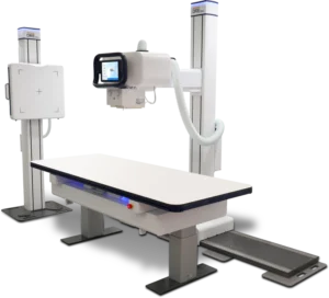
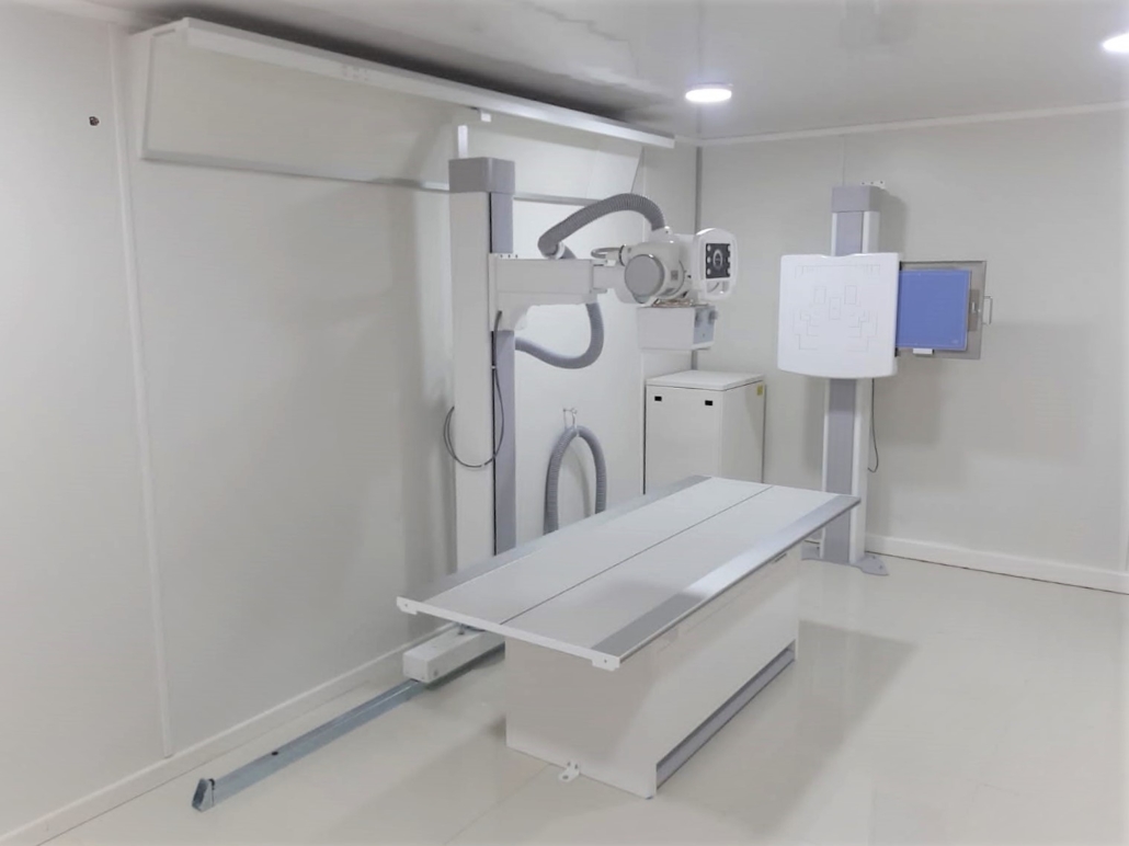
Today, there is a broad range of DR units to fit the needs and budget of every imaging facility. Retrofit DR panels, which come in different sizes and features, enable “Instant DR” by upgrading analog film or CR-based X-ray systems to DR. DR rooms are available with integrated x-ray generators and many possible room configurations. Mobile DR units enable imaging to be done at the patient’s bedside in critical care units or even houses. Analog mobile units can also be retrofitted with DR to extend their useful life.
DR uses flat panel detectors to capture images. First, X-rays are absorbed in a phosphor screen layer, inside the flat panel detector. The X-rays are then converted into visible light. A photodiode converts this light into photo- charges that are collected via the active matrix TFT sensor of the flat panel detector, creating a signal from each pixel. These signals are amplified, digitized, processed, and sent to the acquisition workstation. From the DR acquisition station, they are sent to a display, distribution, and archival system (commonly referred to as a picture archive and communication system, or PACS). The digital image can be displayed on a distant computer or even a tablet and shared with other facilities or patients.
Image processing acquisition software. The software should increase productivity: reducing work for radiology staff. This means little to no manual post-processing, automatic window/level adjustments, automatic electronic masking, and excellent area of interest (AOI) accuracy. Configuration and set-up should be easy: the software should work well out of the box with little or no ongoing maintenance, include simple and understandable adjustment settings, and avoid complex parameter adjustments that require set-up and maintenance by imaging specialists. It should provide consistent performance, for all body types and patient ages, over a wide range of exposure factors. It should not create artifacts, should be tolerant of over and underexposure, and should especially be low-dose friendly.
The importance of anti-scatter grids. The function of the grid is to absorb the scattered radiation coming from the patient before it hits the receptor, therefore increasing image contrast. The anti-scatter grid plays an important role in enhancing image quality in projection radiography by transmitting a majority of primary radiation and selectively rejecting scattered radiation. Grid performance factors can also have a significant impact on the digital image. It is important that the anti-scatter grid is selected correctly for the DR system used, and that the DR software itself can handle eliminating any artifacts that come from the grid, as reciprocating buckys that move the grid should not be used with DR detectors.
Moving from CR to DR can be simple with the right equipment and bring significant improvements and revenue for practices. If you’d like to learn more about how to easily upgrade to digital radiography with JPI, please click here.
*Article extracted from the paper “Moving from CR to DR” by George Curley, Bruce Apgar, Dirk Vandenbroucke.