What is PACS?
A PACS, or picture archiving and communication system, is essentially a medical imaging technology used for storing, retrieving, presenting, and sharing images produced by medical hardware, such as x-ray. Radiology professionals used a PACS to store all diagnostic imaging files. From there, any member of that medical team can quickly search through this information and retrieve images as desired.
Digital Imaging and Communications in Medicine (DICOM) is a protocol that enables the sharing of images and other medical information. DICOM enables PACS technologies. The main purpose of a PACS is to make to not only make the viewing of images easier, but also for better management of important medical data.
PACS technologies have been used in the healthcare industry for decades. To meet this need with today’s technology, JPI Healthcare Solutions developed ExamVue PACS, which is a user-friendly viewing system. In addition to being interoperate with other devices using the DICOM protocol, ExamVue PACS has become well-recognized for its intuitive interface, simple and fast workflow, and convenient diagnostic functions.
With up to 10 viewers, JPI’s ExamVue PACS can be used in both the small clinic and larger practices. This powerful software can be quickly and easily installed throughout your office—both to show your patient’s their x-rays or perform your own diagnoses in your private office. Features include easy image importation, image stitching, comparisons, and specialized ortho, chiro, and veterinary versions, each with their respective specialized tools.
JPI’s ExamVue PACS also offers:
• Flexible Resolution
• Changeable Skin Color (Blue, Pink, Black)
• Support DICOM Q/R, DICOMDIR
• Supports Multiple Viewers
• Supports Dual Monitor Function (Worklist, Viewer)
• DICOM 3.0 Compatibility
Please feel free to contact JPI for more information on our product and software offerings.
How Much Does Digital X-Ray Cost
One of the largest deciding factors in the choice to add or upgrade to digital x-ray imaging is cost. Despite being one of the most frequently asked questions, no imaging solutions provider can tell you exactly how much making changes will add up to without talking to you about your specific needs. Today, most Computed Radiography (CR) solutions have a clinic price below $20,000 while Digital Radiography (DR) solutions are typically priced closer to $30,000. However, there are several factors that commonly affect the cost of making such changes in your business. The three most impact factors are x-ray sourcing, clinic needs, and additional parts.
X-Ray Sourcing
First and foremost, do you already have an x-ray source? This is one of the keys to the question of overall cost. If your clinic does not already have an x-ray source or is in need of new equipment, this will have a significant effect on the cost of adding a digital imaging solution. New x-ray sources can require new wiring and shielding as well so make sure to talk to an experienced imaging solutions provider. You may also want to upgrade an existing x-ray source but, unless the system’s generator has pre-existing problems, this probably isn’t necessary.
Clinic Needs
There are two different image capturing options most clinics consider when adding a digital imaging solution. CR systems are cassette-based digital processing, while DR is a direct capture digital image process. High volume clinics should consider DR for its efficiency, but is also most costly on average, and can lack flexibility compared to a CR system.
Additional Parts
If you choose to go with a DR system, you will also have to decide whether or not you want or need a wireless detector. Many radiology rooms will actually work better with a tethered detector wired directly into your computer, but there are also many applications that demand or benefit from a movable, wireless DR. You may also want to add a Picture Archiving Communication System (PACS) to view your images on other computers, protective or weight-bearing covers for your equipment, and other accessories which can certainly add up.
Ultimately, the individual needs and overall situation of the clinic can determine how moving to a digital imaging solution will cost. It’s important to go into the market for a system knowing these factors will impact the price of your upgrade. While a price for either CR or DR system cannot be easily calculated, knowing these factors when talking to an experienced imaging solutions provider will help ease the buying process and allow you to purchase what best suits your clinic needs.
What Are X-Ray Grids?
The basic purpose of X-ray grid use is to enhance the contrast and quality of the medial image by removing the scatter radiation. With regard to grid specifications, the object that scatter radiation arises is thicker than 10cm. The scatter radiation is also known as “Compton scatter radiation.” Depending on the purpose of radiograph such as chiropractic, veterinary, etc. and the direction of the X-ray, such as AP (anterior to posterior), PA, Lateral, Oblique etc., each grid specification should be different. Also, kVp and mA should be adjusted to acquire better X-ray images through the use of a grid.
Whenever X-rays pass through matter, some of the x-ray photons will interact with the atoms of the matter. When taking an X-ray, may incoming X-ray photons are absorbed by the bone, a high attenuation material, while other tissues absorb less. This produces an image of dark and light on the X-ray film. This effect is like holding up paper to a light to see the writing on the other side. However, absorption is not the end. As part of the absorption, a second photon with less energy will be produced, flying off in a different direction than the photon that was absorbed. These “scattered” photons will create a random grayness on the image, reducing the contrast between body tissues, and making it difficult to read the image clearly.
The are five ways X-rays interact with matter. These include photo electronic (FE), Compton Scattering (C), Pair Production, Tomson Scattering (R), and Photodisintegration (PD). In low energy x-rays below 100keV, only photoelectronic absorption and Compton scattering are significant.
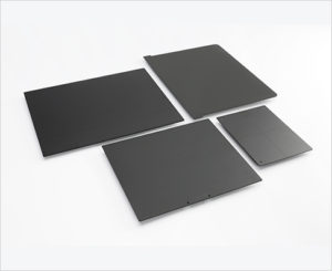
Types of Grids
X-ray grids are divided into four kinds based on their designs. There are parallel types, focused types, crisscross types, and tapered types. A single grid can be more than one type.
Parallel Type Grids
A grid where the absorbing strips are parallel to each other in their longitudinal axis. Most linear grids are also focused (i.e. the strips are slightly tilted, converging at a line in space (the convergent line). A non-focused linear grid has strips that are parallel when viewed in cross-section; this is called a parallel grid. Many X-ray tables are equipped with linear, focused grids, and the strips in these grids are parallel with the long axis of the table, allowing the X-ray tube to be tilted in this direction without changing the effectiveness of the grid.
Focused Type Grids
A grid in which the absorbing strips are slightly angled towards the focal spot. The grid can therefore be used only at a specified focal distance (actually inside a narrow distance interval around the specific distance). Otherwise, the grid will absorb the primary radiation and parts of the film are barely exposed. Focused grids may be linear or crossed.
Criss-Cross Type Grids
A grid consisting of two superimposed parallel grids having the same focusing distance. Such grids are very efficient in removing scattered radiation but must be arranged at exactly right angle to the beam. Th use of such grids is therefore limited.
Tapered Type Grids
A grid that the surface is tapered into the center of grid, functioning similar to a focused grid. All of the strips are parallel to each other, and the tapered surface is toward the focal spot.
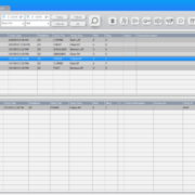
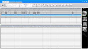
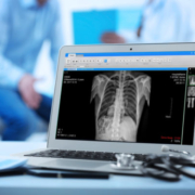
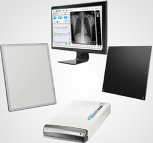
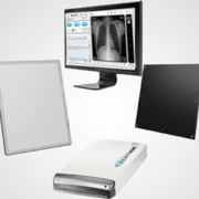
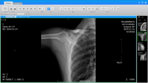
Computed Radiography (CR) vs. Digital Radiography (DR)
Which Should You Choose?
When deciding how to modernize your practice with the use of digital radiography it is important that research be done into which way would be best for your clinic. In the choice between Computed Radiography and Digital Radiography you need to weigh the abilities of each, and the needs you wish to satisfy.
What is Computed Radiography (CR) ?
Computed Radiography — or CR for short — is the use of a Phosphor Imaging Plate to create a digital image. CR uses a cassette based system like analog film and is more commonly considered to be a bridge between classical radiography and the increasingly popular fully digital methods.
Often considered the most initially cost friendly choice, CR is used almost exactly like conventional film, and so requires few changes to your office or workflow, and requires a smaller initial investment compared to DR devices of similar quality. CR systems also do not speed up your workflow in the same way as a DR panel and can require more maintenance. CR cassettes may additionally run the risk of getting damaged if improperly stored or handled, but are much cheaper to replace if dropped than a wireless DR panel.
Pros
- Low initial investment
- Compatible with a wide range of traditional systems
- Effective for smaller or low volume clinics
- Multiple sizes allow for greater flexibility
Cons
- Long time to view image
- Risk of overexposure
- High Maintenance
What is Digital Radiography (DR)?
Digital Radiography (DR) is the latest advancement to the radiography field, using a digital x-ray detector to automatically acquire images and transfer them to a computer for viewing. This system is additionally capable of fixed or mobile use.
While it is the more expensive option, the Digital Radiography system comes to the table with much higher efficiency and quality that more than justifies the price for many users. Given its high volume capabilities it is often the choice for larger or busier clinics.
Pros
- Faster image acquisition
- Better quality images
- High volume capacity
- Greater dose efficiency [for Cesium Iodide (CsI) panels]
Cons
- More expensive initial costs
- Requires protection from dropping or mishandling
- Protection cover is recommended if panel will be removed from the bucky tray or table
If you’d like to learn more about your options for upgrading to digital X-ray, please feel free to email us or call 516-513-1330 ext. 3 to speak with an Account Manager.